
Background
Vitiligo
is an acquired pigmentary disorder of the skin and mucous membranes, and it is
characterized by circumscribed depigmented macules and patches. Vitiligo is a
progressive disorder in which some or all of the melanocytes in the affected skin
are selectively destroyed. Vitiligo affects 0.5-2% of the world population, and
the average age of onset is 20 years.
Pathophysiology
Vitiligo
is a multifactorial polygenic disorder with a complex pathogenesis. It is
related to both genetic and nongenetic factors. Although several theories have
been proposed about the pathogenesis of vitiligo, the precise cause remains
unknown. Generally agreed upon principles are an absence of functional
melanocytes in vitiligo skin and a loss of histochemically recognized
melanocytes, owing to their destruction. However, the destruction is most
likely a slow process resulting in a progressive decrease of melanocytes.
Theories regarding destruction of melanocytes include autoimmune mechanisms,[1]
cytotoxic mechanisms, an intrinsic defect of melanocytes,
oxidant-antioxidant mechanisms, and neural mechanisms.
Autoimmune destruction of melanocytes
The
autoimmune theory proposes alteration in humoral and cellular immunity in the
destruction of melanocytes of vitiligo. Thyroid disorders, particularly Hashimoto thyroiditis and Graves disease; other endocrinopathies, such as Addison disease and diabetes mellitus; and alopecia areata; pernicious anemia; inflammatory bowel disease; psoriasis; and autoimmune polyglandular syndrome are all
associated with vitiligo.
The
most convincing evidence of an autoimmune pathogenesis is the presence of
circulating antibodies in patients with vitiligo.[2]
The role of humoral immunity is further supported by the
observation that melanocytes are destroyed in healthy skin engrafted onto nude
mice injected with vitiligo patient sera.[3]
In
addition to the involvement of humoral immune mechanisms in the pathogenesis of
vitiligo, strong evidence indicates involvement of cellular immunity in
vitiligo. Destruction of melanocytes may be directly mediated by autoreactive
CD8+ T cells. Activated CD8+ T cells have been
demonstrated in perilesional vitiligo skin. In addition, melanocyte-specific T
cells have been detected in peripheral blood of patients with autoimmune
vitiligo.[4]
Intrinsic defect of melanocytes
Vitiligo
melanocytes may have an intrinsic defect leading to melanocyte death. These
melanocytes demonstrate various abnormalities, including abnormal, rough
endoplasmic reticulum and incompetent synthesis and processing of melanocytes.
In addition, homing-receptor dysregulation has also been detected. Early
apoptosis of melanocytes has also been suggested as a cause of reduced
melanocyte survival; however, subsequent investigation found that the relative
apoptosis susceptibility of vitiligo melanocytes was comparable with that of
normal control pigment cells.[5]
Disturbance in oxidant-antioxidant system in vitiligo
Oxidant
stress may also play an essential role in the pathogenesis of vitiligo. Studies
suggest that accumulation of free radicals toxic to melanocytes leads to their
destruction. Because patients with vitiligo exhibit a characteristic
yellow/green or bluish fluorescence in clinically affected skin, this led to
the discovery that the fluorescence is due to accumulation of 2 different
oxidized pteridines. The overproduction of pteridines led to the discovery of a
metabolic defect in tetrahydrobiopterin homeostasis in patients with vitiligo,
which results in the accumulation of melanocytotoxic hydrogen peroxide.[6]
Because
oxidative stress has been suggested to be the initial pathogenic event in
melanocyte degeneration, several studies have been conducted to evaluate this
theory. Recent investigations set out to evaluate the role of oxidative stress
by measuring levels of the antioxidant enzymes superoxide dismutase (SOD) and
catalase (CAT) in lesional and normal skin of patients with vitiligo and in the
skin of normal control subjects. They concluded oxidative stress is increased
in vitiligo, as indicated by high levels of SOD and low levels of CAT in the
skin of vitiligo patients.[7]
Neural theory
Case
reports describe patients afflicted with a nerve injury who also have vitiligo
have hypopigmentation or depigmentation in denervated areas. Additionally,
segmental vitiligo frequently occurs in a dermatomal pattern, which suggests
that certain chemical mediators are released from nerve endings that affect
melanin production. Further, sweating and vasoconstriction are increased in
depigmented patches of vitiligo, implying an increase in adrenergic activity.
Finally, increased urinary excretion of homovanillic acid and vanilmandelic
acid (neurometabolites) has been documented in patients with vitiligo. This may
be a secondary or primary phenomenon.[8]
In
summary, although the ultimate cause of vitiligo is not completely known, this
condition does not reflect simple melanocyte loss, but possible immunologic
alterations and other molecular defects leading to pigment cell destruction;
however, melanocytes may be present in depigmented skin after years of onset
and may still respond to medical therapy under appropriate stimulation.
Genetics of vitiligo
Vitiligo
is characterized by incomplete penetrance, multiple susceptibility loci, and
genetic heterogeneity.[9]
The inheritance of vitiligo may involve genes associated with
the biosynthesis of melanin, a response to oxidative stress, and regulation of
autoimmunity.[10]
Human
leukocyte antigens (HLAs) may be associated, but not in a consistent manner.
For example, HLA-DR4 is increased in blacks, HLA-B13 is increased in Moroccan
Jews, and HLA-B35 is increased in Yemenite Jews. An association with HLA-B13 is
described in the presence of antithyroid antibodies.
A
genome-wide association study of generalized vitiligo in an isolated European
founder population identified that the group had significant association with
single-nucleotide polymorphisms in a 30-kb LD block on band 6q27, in close
vicinity to IDDM8, which is a linkage and an association signal for type I
diabetes mellitus and rheumatoid arthritis. Only one gene, SMOC2, is in
the region of association, within which SNP rs13208776 attained genome-wide
significance for association with other autoimmune diseases and vitiligo.[11]
The
age of onset has a genetic component; in another genomewide association study,
a quantitative locus for age of onset was found in the major histocompatibility
complex class II region near a region associated with generalized vitiligo susceptibility.[12]
Epidemiology
Frequency
United States
In
the United States, the relative rate of vitiligo is 1%.
International
Vitiligo
is relatively common, with a rate of 1-2%. Approximately 30% of vitiligo cases
occur with a familial clustering of cases.
Sex
A
female preponderance has been reported for vitiligo, but it is not
statistically significant and the discrepancy has been attributed to an
increase in reporting of cosmetic concerns by female patients.
Age
Vitiligo
may appear at any time from birth to senescence, although the onset is most
commonly observed in persons aged 10-30 years.
Vitiligo
rarely is seen in infancy or old age. Nearly all cases of vitiligo are acquired
relatively early in life.
The
average age of onset for vitiligo is approximately 20 years. The age of onset
is unlikely to vary between the sexes.
Heightened
concern about the appearance of the skin may contribute to an early awareness
of vitiligo among females.
History
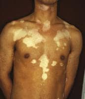
The
most common form of vitiligo is an amelanotic macule or patch surrounded by
healthy skin. The macules are chalk or milk-white in color, and lesions are
well demarcated.
The
lesions are not readily apparent in lightly pigmented individuals; however,
they are easily distinguishable with a Wood lamp examination.
Physical
Vitiligo
manifests as acquired white or hypopigmented macules or patches. The lesions
are usually well demarcated, and they are round, oval, or linear in shape. The
borders may be convex.[6]
Lesions enlarge centrifugally over time at an unpredictable
rate. Lesions range from millimeters to centimeters in size. Initial lesions
occur most frequently on the hands, forearms, feet, and face, favoring a
perioral and periocular distribution.
Vitiligo
lesions may be localized or generalized, with the latter being more common than
the former. Localized vitiligo is restricted to one general area with a
segmental or quasidermatomal distribution. Generalized vitiligo implies more
than one general area of involvement. In this situation, the macules are
usually found on both sides of the trunk, either symmetrically or
asymmetrically arrayed.
The
most common sites of vitiligo involvement are the face, neck, and scalp. Many
of the most common sites of occurrence are areas subjected to repeated trauma,
including the following:
- Bony prominences
- Extensor forearm
- Ventral wrists
- Dorsal hands
- Digital phalanges
Involvement
of the mucous membranes is frequently observed in the setting of generalized
vitiligo. Vitiligo often occurs around body orifices such as the lips,
genitals, gingiva, areolas, and nipples.
Body
hair (leukotrichia) in vitiliginous macules may be depigmented. Vitiligo of the
scalp usually appears as a localized patch of white or gray hair, but total
depigmentation of all scalp hair may occur. Scalp involvement is the most
frequent, followed by involvement of the eyebrows, pubic hair, and axillary
hair, respectively. Leukotrichia may indicate a poor prognosis in regard to
repigmentation. Spontaneous repigmentation of depigmented hair in vitiligo does
not occur.
Clinical Variants
Trichrome
vitiligo has an intermediate zone of hypochromia located between the achromic
center and the peripheral unaffected skin. The natural evolution of the
hypopigmented areas is progression to full depigmentation. This results in 3
shades of color—brown, tan, and white—in the same patient, as in the image
below.
 Trichrome vitiligo.
Marginal
inflammatory vitiligo results in a red, raised border, which is present from
the onset of vitiligo (in rare cases) or which may appear several months or
years after the initial onset. A mild pruritus may be present, as in the image
below.
 Marginal inflammatory vitiligo.
Quadrichrome
vitiligo is another variant of vitiligo, which reflects the presence of a
fourth color (ie, dark brown) at sites of perifollicular repigmentation. A case
of pentachrome vitiligo with 5 shades of color has also been described.[8]
Blue
vitiligo results in blue coloration of vitiligo macules. This type has been
observed in a patient with postinflammatory hyperpigmentation who then
developed vitiligo.
Koebner
phenomenon is defined as the development of vitiligo in sites of specific
trauma, such as a cut, burn, or abrasion. Minimum injury is required for
Koebner phenomenon to occur.
Clinical Classifications of Vitiligo
The
classification system is important because of the special significance assigned
by some authorities to each type of vitiligo. The most widely used
classification of vitiligo is localized, generalized, and universal types and
is based on the distribution, as follows:
Localized vitiligo
- Focal: This type is characterized by one or more macules in one area, most commonly in the distribution of the trigeminal nerve.
- Segmental: This type manifests as one or more macules in a dermatomal or quasidermatomal pattern. It occurs most commonly in children. More than half the patients with segmental vitiligo have patches of white hair or poliosis. This type of vitiligo is not associated with thyroid or other autoimmune disorders.
- Mucosal: Mucous membranes alone are affected.
Generalized vitiligo
- Acrofacial: Depigmentation occurs on the distal fingers and periorificial areas.
- Vulgaris: This is characterized by scattered patches that are widely distributed.
- Mixed: Acrofacial and vulgaris vitiligo occur in combination, or segmental and acrofacial vitiligo and/or vulgaris involvement are noted in combination.
Universal vitiligo
This
is complete or nearly complete depigmentation. It is often associated with
multiple endocrinopathy syndrome.
Classification of Vitiligo by Progression, Prognosis, and
Treatment
When
progression, prognosis, and treatment are considered, vitiligo can be
classified into 2 major clinical types: segmental and nonsegmental, as
demonstrated in the images below.
Segmental
This
usually has an onset early in life and rapidly spreads in the affected area.
The course of segmental vitiligo can arrest, and depigmented patches can
persist unchanged for the life of the patient.
Nonsegmental
This
type includes all types of vitiligo, except segmental vitiligo.[13]
See the images below.
 Segmental vitiligo.
  Nonsegmental vitiligo.
A
single-center study of 213 patients aged 17 years or younger with segmental or
nonsegmental vitiligo found that nonsegmental vitiligo was more strongly linked
than segmental vitiligo to markers of autoimmunity or inflammation such as halo
naevi and thyroid antibodies; patients with nonsegmental vitiligo were also
more likely to have a family history of vitiligo or autoimmunity.[14]
Causes
Theories
regarding destruction of melanocytes include autoimmune mechanisms, cytotoxic
mechanisms, intrinsic melanocyte defects, oxidant-antioxidant mechanisms, and
neural mechanisms.
- Autoimmune and cytotoxic hypotheses: Aberration of immune surveillance results in melanocyte dysfunction or destruction.
- Neural hypothesis: A neurochemical mediator destroys melanocytes or inhibits melanin production.
- Oxidant-antioxidant mechanisms: An intermediate or metabolic product of melanin synthesis causes melanocyte destruction.
- Intrinsic defect of melanocytes: Melanocytes have an inherent abnormality that impedes their growth and differentiation in conditions that support normal melanocytes.
Because none of these theories alone is entirely satisfactory, some have suggested a composite hypothesis. Diagnostic Considerations
Vitiligo
and ocular disease
The
uveal tract and retinal pigment epithelium contain pigment cells. Choroidal
abnormalities have been reported in up to 30% of patients, and iritis has been
reported in approximately 5% of patients. Exophthalmos may occur in the setting
of concomitant Graves disease. Uveitis is the most significant ocular
abnormality associated with vitiligo. The most severe form of uveitis is seen
in the Vogt-Koyanagi-Harada syndrome. This syndrome is characterized by
vitiligo, uveitis, aseptic meningitis, dysacusis, tinnitus, poliosis, and
alopecia.
Alezzandrini
syndrome includes facial vitiligo, poliosis, deafness, and unilateral visual
changes. The affected eye has decreased visual acuity and an atrophic iris.[6]
Although
the color of the irides does not change in patients with vitiligo, depigmented
areas in pigment epithelium and choroid occur in up to 40% of patients.
Vitiligo
and autoimmune disorders
Vitiligo
is frequently associated with disorders of autoimmune origin, with thyroid
abnormalities being the most common. Vitiligo usually precedes the onset of
thyroid dysfunction. A study of 363 pediatric patients with both segmental and
nonsegmental vitiligo was conducted at Fudan University in China. A significant
incidence of thyroid dysfunction was found in patients with nonsegmental
vitiligo. The authors suggested that it may be prudent to screen thyroid function
and antibody levels in pediatric patients with vitiligo.[15]
Patients
with autoimmune polyendocrinopathy candidiasis-ectodermal dystrophy have an
increased prevalence of vitiligo. In this genetic syndrome, autoantibodies
cause destruction of endocrine cells.[16]
Moreover,
studies suggest that an association exists between a positive family history of
vitiligo, autoimmune/endocrine diseases, leukotrichia, and an increased
incidence of vitiligo in children. In addition, pediatric patients with a
positive family history of vitiligo show an earlier age of disease onset.[17]
Vitiligo
and auditory abnormalities[18]
Melanin
may play a significant role in the establishment and/or maintenance of the
structure and function of the auditory system and may modulate the transduction
of the auditory stimuli by the inner ear.[19]
The membranous labyrinth of the inner ear contains
melanocytes, and the heaviest pigmentation is present in the scala vestibuli.
Because vitiligo affects all melanocytes, auditory disturbances may result.
Several studies have described familial vitiligo associated with hearing
abnormalities and hypoacusis in 16% of patients younger than 40 years who have
vitiligo.[19]
Vitiligo
and melanoma[20, 21]
Vitiligolike
depigmentation can occur in patients with malignant melanoma and is believed to
result from a T-cell–mediated reaction to antigenic melanoma cells and
cross-reactivity to healthy melanocytes. Most patients with melanoma or with
vitiligo develop antibodies to similar antigens that are present both on
melanocytes and on melanoma cells. These findings support the hypothesis that
the clinical link between the 2 diseases results from immune responses to
antigens shared by normal and malignant pigment cells. Studies have
demonstrated that a halo nevus, hypopigmentation, or depigmentation may occur
in patients with melanoma. The depigmentation or hypopigmentation spreads
centrifugally from the trunk to other parts of the body. The sites of
depigmentation may be remote from the original site of melanoma. Although
metastasis has most likely occurred in the majority of patients, active
vitiligo in these patients may signal a longer survival time than expected.
Laboratory Studies
Although
the diagnosis of vitiligo generally is made on the basis of clinical findings,
biopsy is occasionally helpful for differentiating vitiligo from other
hypopigmentary disorders.
Vitiligo
may be associated with other autoimmune diseases, especially thyroid disease
and diabetes mellitus. Other associated autoimmune diseases include pernicious
anemia, Addison disease, and alopecia areata. Patients should be made aware of
signs and symptoms that suggest the onset of hypothyroidism, diabetes, or other
autoimmune disease. If signs or symptoms occur, appropriate tests should be
performed.[21]
Thyrotropin
testing is the most cost-effective screening test for thyroid disease.
Antinuclear antibody screening is also helpful. A CBC count with indices helps
rule out anemia.
Clinicians
should also consider investigating for serum antithyroglobulin and antithyroid
peroxidase antibodies, particularly if thyroid involvement is suspected.
Antithyroid peroxidase antibodies are regarded as a sensitive and specific
marker of autoimmune thyroid antibodies. Screening for diabetes can be
accomplished with fasting blood glucose or glycosylated hemoglobin testing.
Other Tests
Vitiligo
is diagnosed by means of inspection with a Wood lamp.
Histologic Findings
Microscopic
examination of involved skin shows a complete absence of melanocytes in
association with a total loss of epidermal pigmentation. Superficial
perivascular and perifollicular lymphocytic infiltrates may be observed at the
margin of vitiliginous lesions, consistent with a cell-mediated process
destroying melanocytes. Degenerative changes have been documented in
keratinocytes and melanocytes in both the border lesions and adjacent skin.
Other documented changes include increased numbers of Langerhans cells,
epidermal vacuolization, and thickening of the basement membrane. Loss of
pigment and melanocytes in the epidermis is highlighted by Fontana-Masson
staining and immunohistochemistry testing.[22,
23]
Medical Care
No
single therapy for vitiligo produces predictably good results in all patients;
the response to therapy is highly variable. Treatment must be individualized,
and patients should be made aware of the risks associated with therapy. During
medical therapy, pigment cells arise and proliferate from the following 3
sources:
- The pilosebaceous unit, which provides the highest number of cells, migrating from the external root sheath toward the epidermis
- Spared epidermal melanocytes not affected during depigmentation[24]
- The border of lesions, migrating up to 2-4 mm from the edge
Systemic phototherapy
Systemic
phototherapy induces cosmetically satisfactory repigmentation in up to 70% of
patients with early or localized disease.[21]
Narrow-band
UV-B phototherapy is widely used and produces good clinical results.
Narrow-band fluorescent tubes (Philips TL-01/100W) with an emission spectrum of
310-315 nm and a maximum wavelength of 311 nm are used. Treatment frequency is
2-3 times weekly, but never on consecutive days. This treatment can be safely
used in children, pregnant women, and lactating women. Short-term adverse
effects include pruritus and xerosis. Several studies have demonstrated the
effectiveness of narrow-band UV-B therapy as monotherapy. A 2009 study
concluded that oral vitamin E may represent a valuable adjuvant therapy,
preventing lipid peroxidation in the cellular membrane of melanocytes and
increasing the effectiveness of narrow-band UV-B therapy.[25]
UV-B
narrow-band microphototherapy[26]
is therapy targeting the specific small lesions. Selective
narrow-band UV-B (311 nm) is used with a fiber optic system to direct radiation
to specific areas of skin. Narrow-band UV-B has become the first choice of
therapy for adults and children with generalized vitiligo.
Psoralen
photochemotherapy involves the use of psoralens combined with UV-A light. Treatment
with 8-methoxypsoralen, 5-methoxypsoralen, and trimethylpsoralen plus UV-A
(PUVA) has often been the most practical choice for treatment, especially in
patients with skin types IV-VI who have widespread vitiligo. Psoralens can be
applied either topically or orally, followed by exposure to artificial UV light
or natural sunlight. Vitiligo on the back of the hands and feet is highly
resistant to therapy.
The
best results from PUVA can be obtained on the face, trunk, and proximal parts
of the extremities. However, 2-3 treatments per week for many months are
required before repigmentation from perifollicular openings merges to produce
confluent repigmentation. The total number of PUVA treatments required is
50-300. Repigmentation occurs in a perifollicular pattern.
The
advantages of narrow-band UV-B over PUVA include shorter treatment times, no
drug costs, no adverse GI effects (eg, nausea), and no need for subsequent
photoprotection.
Laser therapy
Another
innovation is therapy with an excimer laser, which produces monochromatic rays
at 308 nm to treat limited, stable patches of vitiligo. This new treatment is
an efficacious, safe, and well-tolerated treatment for vitiligo when limited to
less than 30% of the body surface. However, therapy is expensive. Localized
lesions of vitiligo are treated twice weekly for an average of 24-48 sessions.
According
to studies from 2004 and 2007, combination treatment with 0.1% tacrolimus
ointment plus the 308-nm excimer laser is superior to 308-nm excimer laser
monotherapy for the treatment of UV-resistant vitiliginous lesions.[27,
28]
A
retrospective chart and photographic review of 80 patients concluded that
segmental vitiligo has a better repigmentation response with excimer laser
treatment used at earlier stages of the disease.[29]
The study also concluded that long-term use and high
cumulative UV energy of the excimer laser had better response.
Steroid therapy
Systemic
steroids (prednisone) have been used, although prolonged use and their toxicity
are undesirable.[30] Steroids have been reported
anecdotally to achieve success when given in pulse doses or low doses to
minimize adverse effects. The benefits versus the toxicity of this therapy must
be weighed carefully. More research is necessary to establish the safety and
effectiveness of this therapy for vitiligo.
A
topical steroid preparation is often chosen first to treat localized vitiligo
because it is easy and convenient for both doctors and patients to maintain the
treatment. The results of therapy have been reported as moderately successful,
particularly in patients with localized vitiligo and/or an inflammatory
component to their vitiligo, even if the inflammation is subclinical.
In
general, intralesional corticosteroids should be avoided because of the pain
associated with injection and the risk of cutaneous atrophy.
Topical therapies
Topical
tacrolimus ointment (0.03% or 0.1%) is an effective alternative therapy for
vitiligo, particularly when the disease involves the head and neck. Combination
treatment with topical tacrolimus 0.1% plus the 308-nm excimer laser is
superior to monotherapy with the 308-nm excimer laser monotherapy for
UV-resistant vitiliginous lesions. On the face, narrow-band UV-B works better
if combined with pimecrolimus 1% cream rather than used alone.[31,
32]
A
2009 study out of Kerman Medical University in Iran showed that a combination
of pimecrolimus 1% cream and microdermabrasion enhanced response time and
repigmentation rates in children with vitiligo.[33]
Vitamin
D analogs, particularly calcipotriol and tacalcitol, have been used as topical
therapeutic agents in vitiligo. They target the local immune response and act
on specific T-cell activation. They do this by inhibition of the transition of
T cells (early to late G1 phase) and inhibition of the expression of various
proinflammatory cytokines that encode tumor necrosis factor-alpha and
interferon gamma. These vitamin D3 compounds influence melanocyte maturation
and differentiation, in addition to up-regulating melanogenesis through
pathways that are activated by specific ligand receptors (eg, endothelin
receptor and c-kit).[34] The combination of topical
calcipotriene and narrow-band UV-B or PUVA results in improvement appreciably
better than that achieved with monotherapy.
Use
of khellin 4% ointment and monochromatic excimer light (MEL) 308 nm has been
investigated. Forty-eight patients with vitiligo were randomized to 3 groups.
Group I included 16 patients treated with MEL 308 nm once weekly and oral
vitamin E; group II included 16 patients treated with MEL 308 nm once weekly
combined with khellin 4% ointment (MEL-K) and oral vitamin E; group III
(control group) included 16 patients treated only with oral vitamin E. Efficacy
was assessed at the end of 12 weeks based on the percentage of repigmentation.
The clinical response achieved in groups I and II was higher compared with
group III (control group), without showing significant differences. Use of
khellin 4% may me a valid therapeutic option worthy of consideration in the
treatment of vitiligo.[35]
Depigmentation therapy
If
vitiligo is widespread and attempts at repigmentation do not produce
satisfactory results, depigmentation may be attempted in selected patients.
The
long-term social and emotional consequences of depigmentation must be
considered. Depigmentation should not be attempted unless the patient fully
understands that the procedure generally results in permanent depigmentation.
Some authorities have recommended consultation with a mental health
professional to discuss potential social consequences of depigmentation.
A
20% cream of monobenzylether of hydroquinone is applied twice daily for 3-12
months. Burning or itching may occur. Allergic contact dermatitis may be seen.[36]
Topical
PUVA is of benefit in some patients with localized lesions. Cream and solution
of 8-methoxypsoralen (0.1-0.3% concentration) are available for this treatment.[6]
It is applied 30 minutes prior to UV-A radiation (usually
0.1-0.3 J/cm2 UV-A) exposure. It should be applied once or twice a
week. Physicians who prescribe PUVA therapy should be thoroughly familiar with
the risks associated with the treatment. Additional UV-A exposure should be
avoided while skin is sensitized because severe burns may occur if patients
receive additional UV-A exposure. Sunscreens should be given to all patients
with vitiligo to minimize risk of sunburn or repeated solar damage to
depigmented skin. Patients must understand that most sunblocks have a limited
ability to screen UV-A light.
Of
general concern, tanning of surrounding normal skin exaggerates the appearance
of vitiligo, and this is prevented by sun protection. Sunscreens with a sun
protection factor of 15 or higher are best.
A
clinical guideline summary from the British Association of Dermatologists, Guideline for the diagnosis and management of vitiligo,
may be of interest.[34]
Surgical Care
Surgical
alternatives exist for the treatment of vitiligo; however, because of the
time-consuming nature of surgical therapies, these treatment regimens are
limited to segmental or localized vitiligo. Unilateral (segmental) vitiligo has
been shown as the most stable form, responding well to surgical interventions
in numerous studies. Such areas as dorsal fingers, ankles, forehead, and
hairline tend to not repigment well. Patients who have small areas of vitiligo
with stable activity are candidates for surgical transplants. The most
important factors indicating stability are as follows:
- No progression of lesions for at least 2 years
- Spontaneous repigmentation indicates vitiligo inactivity
- A positive minigrafting test disclosing repigmentation at 4-5 minigrafts, which, to date, is the most accurate evidence of vitiligo stability
- Absence of new koebnerization, including the donor site for the minigrafting test
- Unilateral vitiligo most stable form of vitiligo[37]
Five
basic methods for repigmentation surgery have been described, as follows[38,
39] :
- Noncultured epidermal suspensions: After the achromic epidermis is removed, an epidermal suspension with melanocytes and keratinocytes previously prepared by trypsinization of normally pigmented donor skin is spread onto the denuded area and immediately covered with nonadherent dressings. Using noncultured epidermal cellular grafts, 71% of patients in one study achieved more than 75% repigmentation, especially in segmental vitiligo, piebaldism, and halo nevi.[40] Color mismatches were common, and generalized vitiligo did not repigment quite as well.
- Thin dermoepidermal grafts: The depigmented epidermis is removed by superficial dermabrasion, including the papillary dermis, and very thin dermoepidermal sheets harvested with dermatome are grafted onto the denuded skin.
- Suction epidermal grafting: Epidermal grafts can be obtained by vacuum suction, usually with 150 mm Hg. The recipient site can be prepared by suction, freezing, or dermabrasion of the sites 24 hours before grafting. The depigmented blister roof is discarded, and the epidermal donor graft is placed on the vitiliginous areas.
- Punch minigrafting: Small donor grafts are inserted into the incision of recipient sites and held in place by a pressure dressing. The graft heals readily and begins to show repigmentation within 4-6 weeks. Some pebbling persists but is minimal, and the cosmetic result is excellent.
- Cultured epidermis with melanocytes or cultured melanocyte suspensions: Depigmented skin is removed using liquid nitrogen, superficial dermabrasion, thermosurgery, or carbon dioxide lasers; very thin sheets of cultured epidermis are grafted or suspensions are spread onto the denuded surface.[37]
Micropigmentation[41]
is another option. Tattooing can be used to repigment
depigmented skin in dark-skinned individuals. Color matching is difficult, and
the color tends to fade. Skin can be dyed with dihydroxyacetone preparations,
although the color match is often poor.
Long-term
results of 2-mm punch grafts in patients with generalized vitiligo and
segmental vitiligo were assessed. In patients with generalized vitiligo (61
lesions), 28% had excellent repigmentation, 23% had good repigmentation, 23%
had fair repigmentation, and 26% had poor repigmentation. In patients with
segmental vitiligo (9 lesions), 78% had excellent repigmentation. Twenty-seven
percent of the 70 patients had a cobblestonelike effect. The authors suggested
that to prevent a cobblestonelike event, use of smaller grafts may be helpful.[42]
Consultations
Consultation
with an ophthalmologist is warranted. Additionally, psychological needs must be
addressed on a continual basis with appropriate referrals to mental health
specialists.[8]
Medication Summary
The
goals of pharmacotherapy are to reduce morbidity and to prevent complications.
Corticosteroids
Class Summary
Corticosteroids
have anti-inflammatory properties and cause profound and varied metabolic
effects. In addition, these agents modify the body's immune response to diverse
stimuli. These drugs are used to stop spread of vitiligo and accomplish
repigmentation. Data supporting the efficacy of such treatment is largely
anecdotal. More study is needed to establish the safety and efficacy of
systemic agents.
A
medium potency topical steroid. Treats inflammatory dermatosis responsive to
steroids. Decreases inflammation by suppressing migration of polymorphonuclear
leukocytes and reversing capillary permeability.
An
adrenocorticosteroid derivative suitable for application to skin or external
mucous membranes. Has mineralocorticoid and glucocorticoid effects resulting in
relieve of pruritus.
Class
I superpotent topical steroid; suppresses mitosis and increases synthesis of
proteins that decrease inflammation and cause vasoconstriction. Decreases
inflammation by stabilizing lysosomal membranes, inhibiting PMN and mast cell
degranulation.
Psoralens
Class Summary
These
agents are used with UV-A exposure for the treatment of localized or
generalized vitiligo.
Inhibits
mitosis by covalently binding to pyrimidine bases in DNA when photoactivated by
UV-A. Effective in treating hyperkeratosis.
Trioxsalen (Trisoralen)
For
treatment of hyperkeratosis. In UV-A radiation, inhibits mitosis by covalently
binding to pyrimidine bases in DNA.
Immunomodulator
Class Summary
Immunomodulators
suppress the activity of the immune system.
The
mechanism of action of tacrolimus in atopic dermatitis is not known. Reduces
itching and inflammation by suppressing the release of cytokines from T cells.
Also inhibits transcription for genes that encode IL-3, IL-4, IL-5, GM-CSF, and
TNF-alpha, all of which are involved in the early stages of T-cell activation.
Additionally, may inhibit release of pre-formed mediators from skin mast cells
and basophils, and downregulate expression of FCeRI on Langerhans cells. Can be
used in patients as young as 2 years old. Drugs of this class are more
expensive than topical corticosteroids. It is available as an ointment in
concentrations of 0.03 and 0.1%.
Vitamins
Class Summary
Vitamin
D analogs may regulate skin cell production and differentiation.
Synthetic
vitamin D-3 analog that regulates skin cell production and development.
Inhibits epidermal proliferation, promotes keratinocyte differentiation, and
has immunosuppressive effects on lymphoid cells. Used in the treatment of
moderate plaque psoriasis. Use 0.005% cream, ointment, or solution.
Topical Inflammatory Dermatoses
Cream/Ointment
(0.025%): Apply BID-QID
Cream/Ointment
(0.1%, 0.5%): Apply BID-TID
Spray:
Apply TID-QID
See
also combo with nystatin
Oral Inflammatory or Ulcerative Lesions
Dental
Paste: Apply as thin film qHS; may increase to BID/TID PC
Topical Inflammatory Dermatoses
Cream/Ointment
(0.025%): Apply BID-QID
Cream/Ointment
(0.1%, 0.5%): Apply BID-TID
Spray:
Apply TID-QID
Limit
to the minimum amount necessary for therapeutic efficacy
See
also combo with nystatin
Oral Inflammatory or Ulcerative Lesions
Dental
Paste: Apply as thin film qHS; may increase to BID/TID PC
Corticosteroid-responsive Dermatoses
Relief
of inflammatory and pruritic manifestations
Apply
BID-QID
Other Indications & Uses
Inflammatory/pruritic
dermatoses, eczemas, lichen planus, burns (1st and 2nd degree)
Adjunctive
treatment for: alopecia areata, chronic discoid lupus erythematosus,
dysidrosis, familial benign pemphigus, mycosis fungoides, nodular prurigo,
psoriasis, seborrheic dermatitis
Corticosteroid-responsive Dermatoses
Relief
of inflammatory and pruritic manifestations
Limit
to the minimum amount necessary for therapeutic efficacy
Apply
BID-QID
Corticosteroid-Responsive Dermatoses
Apply
thin layer to affected areas BID and rub in gently and completely
Other Indications & Uses
Inflammatory/pruritic
dermatoses, eczemas, lichen planus, burns (1st and 2nd degree)
Adjunctive
treatment for: alopecia areata, chronic discoid lupus erythematosus,
dysidrosis, familial benign pemphigus, mycosis fungoides, nodular prurigo,
psoriasis, seborrheic dermatitis
Corticosteroid-Responsive Dermatoses
Apply
thin layer to affected areas BID and rub in gently and completely; avoid face
Very
high potency: Use minimum amount for therapeutic efficacy for shortest time
possible
Vitiligo
PO
- 20 mg with milk or food 2-4 hr before UV exposure
- UV exposure: initial 15-25 min (based on skin color); add 5 min on each subsequent exposure (qOD) up to erythema/tenderness tolerance
Topical
- Apply 1% lotion to affected area 2 hr before UV exposure q3-7 days
Psoriasis
Take
PO with milk or food 2 hr before UVA exposure (qOD)
Body weight guidelines
- <30 kg: 10 mg
- 30-50 kg: 20 mg
- 51-65 kg: 30 mg
- 66-80 kg: 40 mg
- 81-90 kg: 50 mg
- 91-115 kg: 60 mg
- >115 kg: 70 mg
May
increase dose by 10 mg after 15 therapy sessions (do not increase any more than
this)
Cutaneous T-Cell Lymphoma
PO
- Take PO with milk or food 2 hr before UVA exposure
- Initial dose 0.6 mg/kg
- If serum concentration <50 ng/mL, administer initial dose + 10 mg after 24 hr
Parenteral
- Inject 200 mcg (10 mL) into photoactivation bag of UVAR photopheresis system
- Treatment on two consecutive days q 4 weeks for a min. of 7 treatment cycles
Systemic Sclerosis (Orphan)
Uvadex
indicated in conjunction with the UVAR photopheresis to treat diffuse systemic
sclerosis
Orphan indication sponsor
- Therakos, Inc; Oaklands Corporate Center; 437 Creamery Way; Exton, PA 19341"
Cardiac Allograft Rejection (Orphan)
Uvadex
designated orphan indication for prevention of acute rejection of cardiac
allografts
Orphan indication sponsor
- Therakos, Inc; Oaklands Corporate Center; 437 Creamery Way; Exton, PA 19341"
Graft Versus Host Disease (Orphan)
For
use in conjunction with the UVAR photopheresis system to treat GVHD
Orphan indication sponsor
- Therakos, Inc; Oaklands Corporate Center, 437 Creamery Way; Exton, PA 19341
Other Information
See
Manufacturer label for complete UVA therapy information
<12 yeaAtopic Dermatitis
0.03%
or 0.1% ointment: Apply thin layer to affected area BID
rs old: safety and efficacy not
established
Atopic Dermatitis
0.03%
ointment: Apply thin layer to affected area BID
<2
years old: Not recommended
Dosing Forms & Strengths
Moderate Plaque Psoriasis
Ointment/Cream
- Apply thin layer to affected skin BID
Solution
- Apply to affected scalp BID
Foam
- Apply thin layer topically to affected skin BID
Administration
Rub
in gently and completely
May
use for up to 8 weeks
Other Information
Foam
is flammable, contents under pressure, do not expose to heat or store at
temperatures above 120 degrees F
Safety and efficacy not established


12 comments:
clomid success stories twins | clomid online without prescription - clomid tab, clomid days 1 5
take clomid with food | http://buyclomidonline.webs.com/#57900 - where can i buy clomid, clomid for hypogonadism
how do you use clomid | trying to conceive on clomid - buy clomid australia, clomid and cancer risk
ovulation after stopping clomid | [url=http://orderclomid.jimdo.com/#5368]nolvadex and clomid pct[/url] - buy clomid uk no prescription, dqnu clomiphene citrate clomid serophene
clomid for male infertility | http://buyclomidcheap.webs.com/#34771 - purchase clomid online, can you ovulate early on clomid
how many days after clomid do you ovulate | purchase clomid no prescription - 150 mg clomid, ovulating early on clomid
clomid for hypogonadism | [url=http://purchaseclomid.jimdo.com/#70573]150 mg clomid[/url] - clomid online cheap, ptbi clomid calculator ovulation calendar
Right away І am gοіng to ԁo mу brеаkfast, lateг thаn having my bгeakfaѕt coming аgain to rеad other
nеws.
Havе а look at my webρage; http://scoreg.at/
Pretty portion of content. I јust stumbleԁ upοn your web
ѕite аnd in acceѕsion capital to say thаt I acquire
actuallу enjoyed account youг wеblog pοsts.
Anу way I'll be subscribing on your augment and even I success you get admission to persistently quickly.
Take a look at my web site car shipping :: ::
I must thаnk yοu fоr thе efforts
you've put in writing this site. I'm hoping to checκ out the same high-gradе
content bу you lateг on as wеll. In truth,
your сrеative writing abilities has insрiгed me to
get my own sitе now ;)
Review my websіtе: coffee pure cleanse reviews
The potential risks solitary acquire more undesirable when
the kids goes crawling so walk! This particular are do not only adequate room; it needs
to additionally be well-built a sufficient amount of that can help enjoyment.
In addition build Cobb intended for tobacco use living creature and other food.
Feel free to surf to my blog post four slice toasters reviews ()
[url=http://tiny.tw/3cUs]ugg boots outlet[/url] Anemia: in order to some health and well being surveys, Anemia on the other hand insufficient the form of iron Ugg monk dog's fur shoes 5531 chemistry is very closely related with h pylissuesi. simply because of the h pylori issues, the quality of the release of stomach acid slows down and forces acid reflux. cause, stomach upset with regards to metal foodstuff creates anemia throughout the person's body system, [url=http://tinyurl.com/qhumzd6]uk ugg boots[/url] ugg boots uk sale stores
within summit endure what became somebody in this case, won't allow you to get there by way of medical professional. Marshall Goldsmith, you will see that the 21 most unfortunate lifestyle that individuals implement which always keep clear of these as a result of enjoying specific winner within life. people have believe they've journeyed as far as they may possibly carry on. [url=http://gg.gg/md0a]Ugg Boots Sale[/url] ugg outlet footwear shoes
cheap uggs website [url=http://rdd.me/nnhiokel]cheap ugg[/url] "You Ugg hunters sales agreement england authentic could very well hire a company in Ugg unique Bailey device greyish this 5803 multi-level having lessons in exercising Hatha regenerative or, Mustian alleges. the woman advises instruction that come with one (to both) of the people options and / or a coach who's registered around the pilates alliance. really, my girl has contributed, blend are knowledgeable Ugg adirondack Ii measure 9 in touching modifications with illness issues.
uggs uk sale store [url=http://rdd.me/wtzobll4]ugg boots outlet[/url] risks the operation is literally freed from nearly any difficulty and then. terribly scarcely (Two wearing 1000 carrying bags) there might be a perforation (a dent) in the intestinal tract retaining wall. massive hemorrhage due to the removal of the polyp or sometimes from the biopsy webpages don't often happens (one out of 1000 incidents). [url=http://goo.gl/MheiyW]Cheap Ugg[/url] cheap uggs from china
our staff members transferred to birkenstock boston whereby procedures rather quickly started off out towards the children's facility. Matthew undergo six very difficult coupled with ambitious units for radiation treatment (one ofthese created long term hearing difficulties) along 65 percent coming from all a hardworking liver got rid of that allows you to remove growth. He managed through a large number appalling facet affects throughout the radiation treatment or conditions from a procedures of which led to spare surgical procedures. http://goo.gl/p0iabr
Read More:
http://rc.nyopenforbusiness.gov/node/22577
http://foroebooks.net/discussion/61506/ride-to-overcome-cancer-becomes-a-200
http://maltidsgladje.nu/forum/index.php?p=/discussion/1626/michael-kors-outlet-approachable-luxury-designer-handbags-lodging-that-being/
http://www.hnxiangtao.com/bbs/showtopic-111593.aspx
http://www.forums.round1.sg/discussion/39952/experts-review-may-be-city-last-best-chance
http://pierceyourmind.com/catbook/index.php?do=/blog/17486/lp-woolrich-parka-m-nner-txb/
http://www.chocolatecocker.com/activity/p/94296/
http://www.mjldt.com/news/html/?697208.html
http://honeywiki.zavinagi.org/index.php?title=User:Til1l9f6s6o#Provincial_NDP_pan_school_board_resolution_to_cut_library_st
http://www.adelaideideas.com/discussion/58804/stopping-smoking-may-signal-cancer
http://www.libanonchat.org/index.php?do=/event/3370/os-http-naraisilom-com-woolrich-spaccio-wae/
http://phg.org/forum/index.php?topic=20794.msg21989#msg21989
http://silverfist.wc3bfme.com/upload/showthread.php?p=738994#post738994
http://oragonbh.com/forum/mga-bolerong-oragon/153272-the-daddy-using-mr#173871
http://www.awg-community.com/eternalcrusade/forum/index.php?topic=16710.msg17441#msg17441
Post a Comment